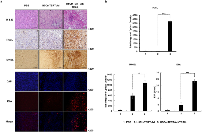
Figure 6.
Histological and immunohistochemical analyses of tumour tissues treated with oncolytic adenoviruses. U87MG tumours established in nude mice were treated with PBS, H5CmTERT-Ad or H5CmTERT-Ad/TRAIL on days 1, 3, and 5, and the tumours were harvested on the 3rd day after administration of the final treatment. (a) Representative sections were stained with hematoxylin and eosin (H & E). The expression levels of TRAIL and E1A were assessed by immunohistochemical analysis. A TUNEL assay was performed to detect apoptosis. Data are representative of three independent experiments. Original magnification: ×200 and ×400. (b) Semi-quantitative analysis of TRAIL-, TUNEL-, or E1A-stained sections in the MetaMorph image analysis software or ImageJ software. All the results are shown as means ± SD; **P < 0.01, ***P < 0.001.

