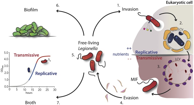
Figure 1.
Schematic overview of the L. pneumophila morphological states during its growth cycle. 1. Uptake of virulent L. pneumophila by the host cell like protozoa or macrophages through convention or coiling phagocytosis. 2. After internalization, the bacteria evade the phagosome-lysosome fusion and start the intracellular multiplication within the LCV, which is surrounded by vesicles (in yellow) rich in lipids and proteins. 3. Nutrient starvation induces the activation of the stringent response and morphological changes. Bacteria express the transmissive traits such as motility (flagella) and become cytotoxic. 4. These infectious bacteria are able to lyse the vacuolar membrane and are released in the extracellular environment. 5. Free-living transmissive bacteria may start a new cycle or persist in the extracellular environment as planktonic form. 6. Alternatively, L. pneumophila may be associated within biofilms, either in natural fresh-water habitats or artificial ones. 7. In broth culture, L. pneumophila displays also a biphasic life cycle, which closely mimics the replicative and transmissive intracellular forms.

