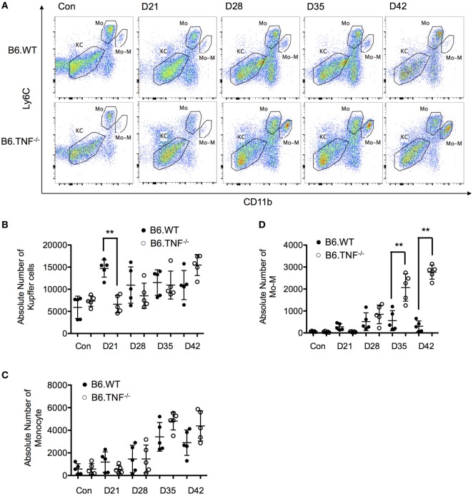
Figure 4.
Analysis of resident and inflammatory myeloid populations in the liver in Leishmania major infected B6.WT and B6.TNF−/− mice. (A) Flow cytometry analysis was used to demonstrate the presence of three different liver macrophage populations based on the markers CD45, F4/80, CD11b, and Ly6C from B6.WT and B6.TNF−/− mice and to analyze the changes of these populations over the course of L. major BNI infection. Kupffer cells (KCs) were defined as CD45+F4/80+CD11b−Ly6C−, inflammatory monocytes (Mo) as CD45+F4/80+CD11blowLy6Chi and monocyte-derived macrophages (Mo-M) as CD45+F4/80+CD11bhiLy6Clow. A representative staining is shown. Quantification by flow cytometry of the total populations of (B) KC, (C) Mo, and (D) Mo-M from five B6.WT and B6.TNF−/− mice in the course of L. major BNI infection is shown. Each error bar represents means ± SD from one experiment. Results were confirmed by two independent experiments. The p-values were calculated using two tailed Mann–Whitney U-test (**p < 0.01).

