
Figure 8.
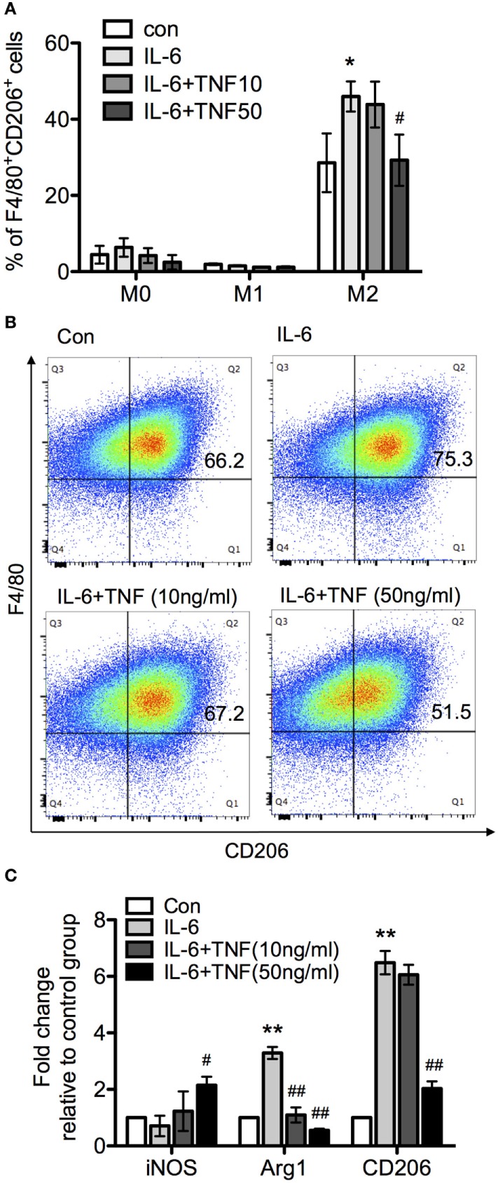
The effect of tumor necrosis factor (TNF) and IL-6 in macrophage differentiation. Bone marrow-derived macrophages were cultivated in the presence of macrophage colony-stimulating factor and harvested after 8 days. Subsequently, macrophages were exposed to LPS and interferon-γ or IL-4 for 24 h to generate M1 and M2 macrophages. The upregulation of the marker molecules F/80 and CD206 was quantified. Unchanged macrophages were considered as M0 phenotype. (A) M0, M1, and M2 macrophages were incubated with IL-6 or IL-6/TNF, and differentiation was analyzed. All the data are presented as means ± SD after normalization to control group values of three experiments. The p-values were calculated using one-way ANOVA and Tukey’s comparison test (*p < 0.05 when compared to the control group, and #p < 0.05 when comparing to the IL-6-treated group). (B) Flow cytometry of cells from each culture condition using the macrophage markers F4/80 and CD206. (C) Quantification of the expression of inducible nitric oxide synthase (iNOS), arginase-1 (Arg-1), and CD206 in M2 macrophages treated with IL-6 and TNF using qPCR. Results are representative of three independent experiments, and presented as mean ± SD normalized with regard to the control group. The p-values were calculated using one-way ANOVA and Tukey’s comparison test (**p < 0.01 when compared to control group, and #p < 0.05, ##p < 0.01 when compared to the IL-6-treated group).