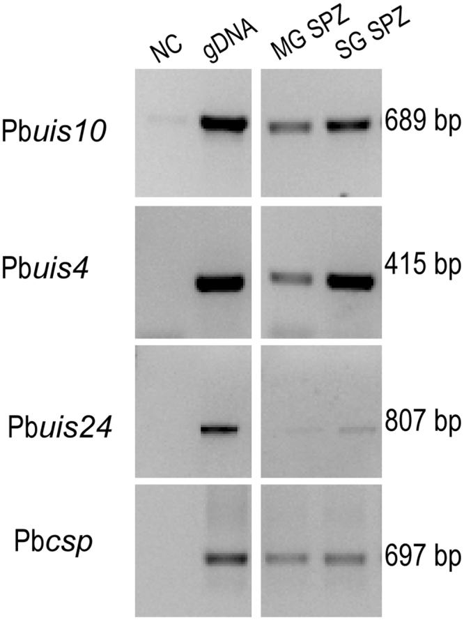
Figure 2.

RT-PCR analysis of P.berghei Pbuis4, Pbuis10, Pbuis24 and Pbcsp transcripts. Templates were derived from gDNA (lane 2), dissected midguts at 11 d pbm sporozoites (lane 3, MG SPZ) and from dissected salivary glands at 21 d pbm (lane 4 SG SPZ). Lane 1 is the negative control without template (NC). The samples were amplified for 35 cycles. Molecular weight of the amplified fragments is indicated to the right. Unprocessed images of the agarose gels are shown in Supplementary Fig. S4b.
