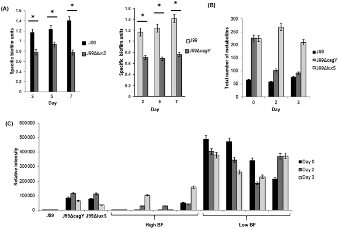
Figure 4.
(A) Specific biofilm unit of wild-type H. pylori J99, J99ΔluxS and J99ΔcagY determined by crystal violet assay. (B) Comparison of number of metabolites detected by LC-MS per sample for H. pylori J99, J99ΔluxS and J99ΔcagY. *Represents p-value < 0.05 comparing between wild-type and mutants. (C) Comparing the relative abundance of PI-Cer(t18:0/18:0(2OH)) in H. pylori J99, J99ΔluxS, J99ΔcagY, high biofilm-forming clinical strains and low biofilm-forming strains.

