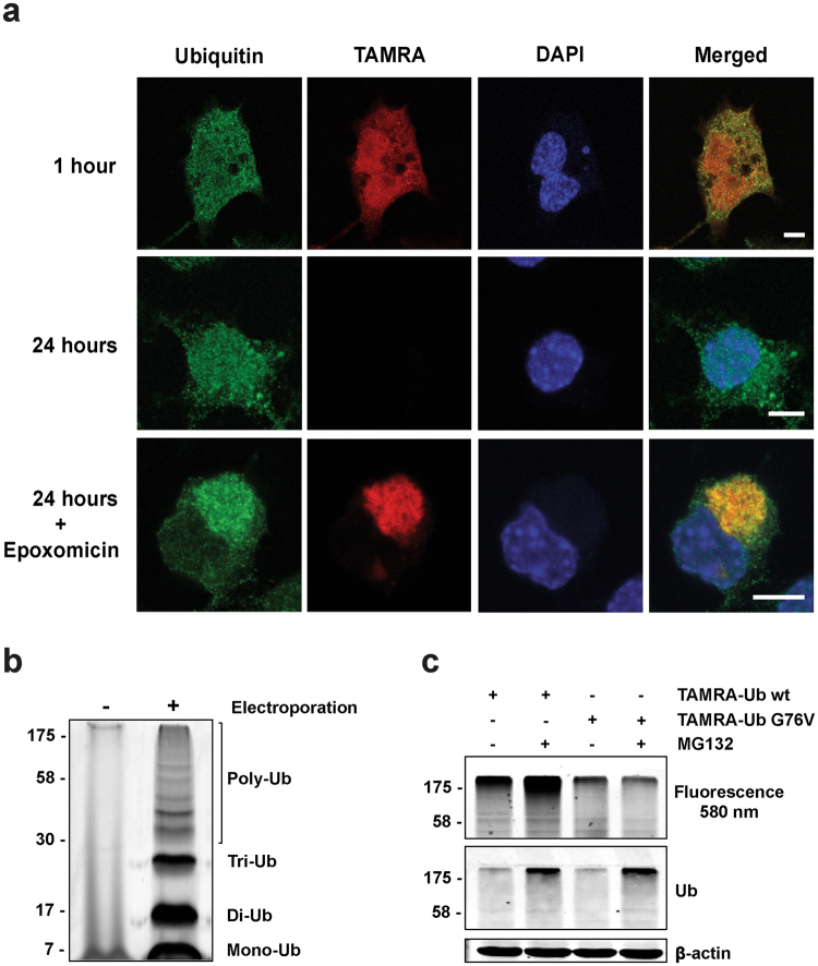
Figure 1.
TAMRA-Ub behaves like endogenous Ub. (a) Confocal images of intracellular ubiquitin localization in Neuro-2A cells one and 24 hours after TAMRA-Ub electroporation. One hour after electroporation cells were treated with 50 nM epoxomicin and stained with an anti-Ub antibody after fixation. Nucleus was stained with DAPI. Scale bar: 5 µm. (b) Neuro-2A cell lysates were harvested two hours after electroporation of TAMRA-Ub and loaded on a SDS-PAGE gel. Fluorescent scan shows TAMRA-Ub incorporated in the poly-Ub tree. (c) Fluorescence scan and immunoblot of SDS-PAGE loaded with cell lysates of Neuro-2A cells electroporated with TAMRA-Ub wildtype (wt) or mutant TAMRA-Ub G76V. One hour after electroporation cells were treated with 20 µM MG132 for additional two hours. Ubiquitinated proteins were detected by anti-Ub antibody. β-actin was used as a loading control.

