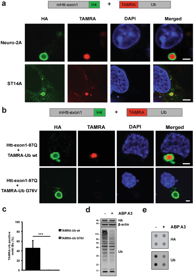
Figure 2.
TAMRA-Ub is recruited into Htt IBs. (a) Confocal images of Htt-exon1-97Q-H4 protein expressed in transient transfected Neuro-2A cells and stable ST14A cells. 24 hours after transfection cells were electroporated with TAMRA-Ub and incubated for additional 24 hours. Fixed cells were immuno-stained with anti-HA antibody and nuclei were stained with DAPI. Scale bar: 3 µm. (b) Confocal images of Neuro-2A cells expressing wild type Htt-exon1-97Q-H4. Cells were electroporated with TAMRA-Ub wild type and the mutant variant G76V 24 hours after transfection and incubated for additional 24 hours. After fixation cells were stained with an anti-HA antibody and the nuclei were stained with DAPI. Scale bar: 2 µm. (c) Quantification of TAMRA-Ub positive IBs. Neuro-2A cells expressing Htt-exon1-97Q-C4 were electroporated with TAMRA-Ub wt and the mutant G76V 24 hours after transfection and were incubated for additional 24 hours. Cells were stained with FlAsH and fixed for microscopic analysis. The percentage of TAMRA-Ub and mutant TAMRA-Ub G76V positive Htt IBs was determined ***p < 0.001, (n = 180). Means and SD are shown. (d) Western blot of Neuro-2A cells expressing Htt-exon1-97Q-H4 were treated with the E1 inhibitor ABP A3 for 6 hours. Ubiquitinated material was stained with the anti-ubiquitin antibody. β-actin was used as a loading control. (e) Filter trap assay (doublets) of mHtt aggregates from Neuro-2A cells transient transfected with the Htt-exon1-97Q-H4 construct for 48 hours. ABP A3 exposure reduces the Ub moiety on SDS-insoluble mHtt aggregates detected by anti-HA and anti-Ub antibodies.

