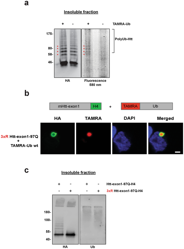
Figure 3.
Aggregated mHtt is conjugated with TAMRA-Ub. (a) Insoluble fraction of Neuro-2A cell lysate 48 hours after transient transfection with Htt-exon1-97Q-H4. After 24 hours expression cells were electroporated with TAMRA-Ub and incubated for additional 24 hours. Formic acid-dissolved inclusion bodies were loaded on a SDS-PAGE and TAMRA signal of ubiquitinated mHtt (asterisks) was detected by fluorescence scan. mHtt proteins were detected on Western blot by anti-HA immunostaining. Non-electroporated cells expressing Htt-exon1-97Q-H4 were used as a control. (b) Confocal images of Neuro-2A cells expressing the mutant 3xR Htt-exon1-97Q-H4. Cells were electroporated with TAMRA-Ub wild type 24 hours after transfection and incubated for additional 24 hours. After fixation cells were stained with an anti-HA antibody and the nuclei were stained with DAPI. Scale bar: 2 µm. (c) Insoluble fractionation of Neuro-2A cell lysate 48 hours after transient transfection with Htt-exon1-97Q-H4 or 3XR Htt-exon1-97Q-H4. Formic acid-dissolved inclusion bodies were loaded on a SDS-PAGE. Htt proteins and ubiquitinated material were detected on Western blot by anti-HA and anti-Ub immunostaining.

