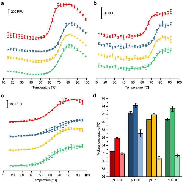
Figure 2.
Differential scanning fluorimetry of β-amylase using different dyes and devices. (a) Melting curves obtained using 1 µM β-amylase with 10× SYPRO Orange in combination with a RT-PCR machine (data points recorded every 1 °C, but 3 °C steps shown for illustration purposes). (b) The same measurement as in a using the microcontroller based device. (c) CPM-fluorescence of 1 µM β-amylase measured with the microcontroller based device. (d) Comparison of the melting temperatures obtained from (a) (dark colours), (b) (medium colours) and c (light colours). The buffers were set to pH 5.0 (red), pH 6.0 (blue), pH 7.0 (yellow) and pH 8.0 (green).

