Fig. 2.
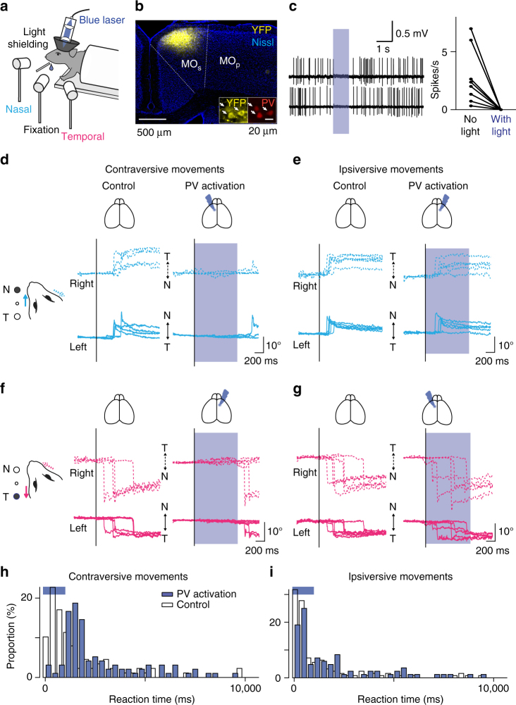
Optogenetic suppression of the MOs during the visually guided eye movement task. a The experimental design. b ChR2 was expressed virally in PV interneurons in Pvalb-IRES-Cre transgenic mice. AAV-DIO-ChR2-eYFP was injected into the MOs (coronal section stained with Nissl), and ChR2-eYFP expression was confined to cells expressing PV (white arrows). c Firing was suppressed during blue light illumination. Left, two representative neurons are shown. Right, blue light illumination completely suppressed firing activity in all eight neurons (p < 0.01, Wilcoxon signed-rank test). The activity was monitored with blind cell-attached recording. d–g Optogenetic suppression of the MOs during the task. The virus was injected into the MOs either in the left (d, g) or right (e, f) hemisphere. Unilateral suppression of the MOs severely impaired eye movements in the contraversive (d, f) but only mildly in the ipsiversive (e, g) direction. h, i Reaction times for the eye movements. PV activation delayed the onset of contraversive eye movements (h, PV activation, n = 96 trials; control, n = 88 trials; p < 10−11), but showed only minor effects on ipsiversive movements (i, PV activation, n = 84 trial; control, n = 126 trials; p = 0.0501, Person’s chi-square test)

