Fig. 6.
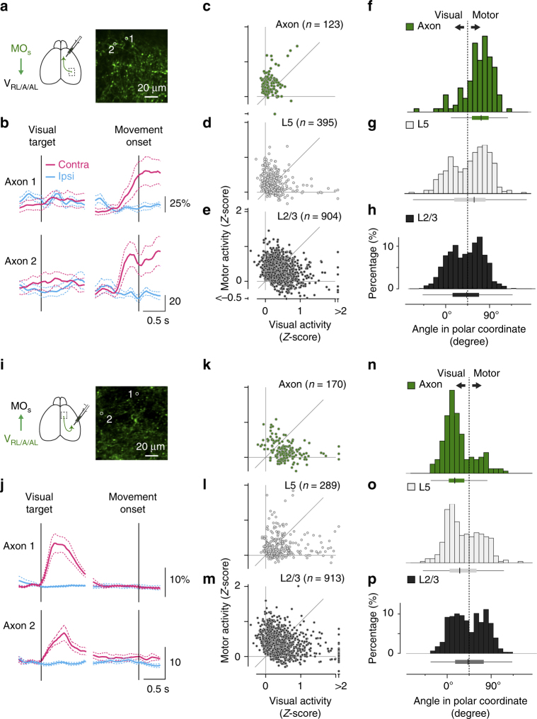
Biased neural coding in MOs and VRL/A/AL axons. a–c Coding in MOs axon terminals projecting to the VRL/A/AL. a GCaMP6f was expressed in MOs neurons, and their axons were imaged in the VRL/A/AL. b Calcium traces from two representative axons (mean ± s.e.m., cyan for the ipsiversive movement condition; magenta for the contraversive). The corresponding ROIs are shown in a. c–e Comparison between visual and motor activity for MOs axons (c, n = 123), L5 somas (d, n = 395), and L2/3 somas (e, n = 904). f–h Histograms of the angle in polar coordinates for MOs axons, L5 neurons, and L2/3 neurons. The distribution of the axons is more biased towards 90 degrees, indicating higher motor selectivity. Median, 25% quantile, and 75% quantile are represented as box plots, whisker length is 1.5 of the interquartile range. i–p Similar to a–h, but for VRL/A/AL axon terminals projecting to the MOs. VRL/A/AL axons, n = 170; VRL/A/AL L5 somas, n = 289; L2/3 somas, n = 913. Note skewed distribution of the axons towards 0 degrees, indicating higher visual selectivity. The histograms for L2/3 somas (h, p) are the same with those in Fig. 5h, j

