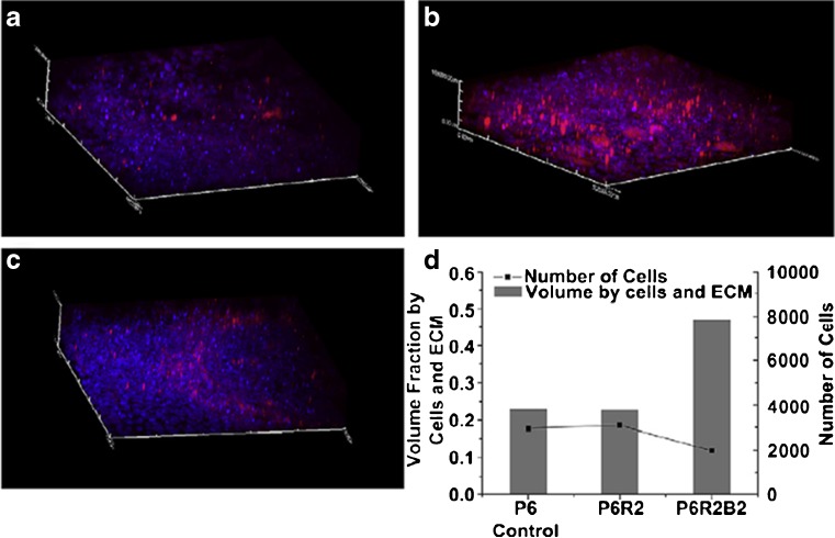
Fig. 12.
3D bone tissue model. a–c Two-photon microscopic images of the 3D bone tissue structure under the influence of the antibiotic- and calcium-eluting micropattern: a P6 control [pure poly(D,L-lactic-co-glycolic) acid (PLGA)]; b PGR2 (6% PLGA, 2% rifampicin); c PGR2B2 (6% PLGA, 2% rifampicin, 2% biphasic calcium phosphate (BCP)). d Quantification of the volume fraction of tissue (left axis) and number of cells (right axis) of three conditions. The presence of BCP in the PGR2B2 condition substantially increased the production of mineralised ECM. The antibiotic did not adversely affect osteoblast viability and tissue formation. Adapted from Lee et al. [123] with permission from Elsevier

