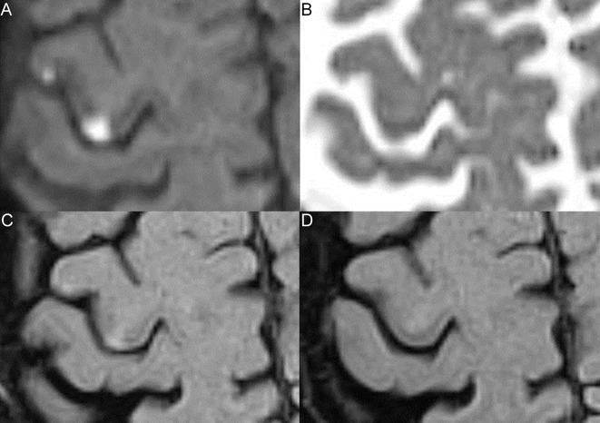
Figure 1.
Lesions with and without 8-week infarction signs. Two cortical grey matter lesions are shown on initial diffusion-weighted imaging (DWI) (A), apparent diffusion coefficient (ADC) (B), initial fluid-attenuated inversion recovery (FLAIR) (C) and 8-week FLAIR (D). (A) Both lesions are DWI-positive. (B) The medial lesion is ADC-confirmed, the lateral lesion shows no ADC-confirmation. (C) Both lesions are initially FLAIR-positive. (D) The medial lesion is 8-week FLAIR-positive, the lateral lesion is 8-week FLAIR-negative.

