Figure 5.

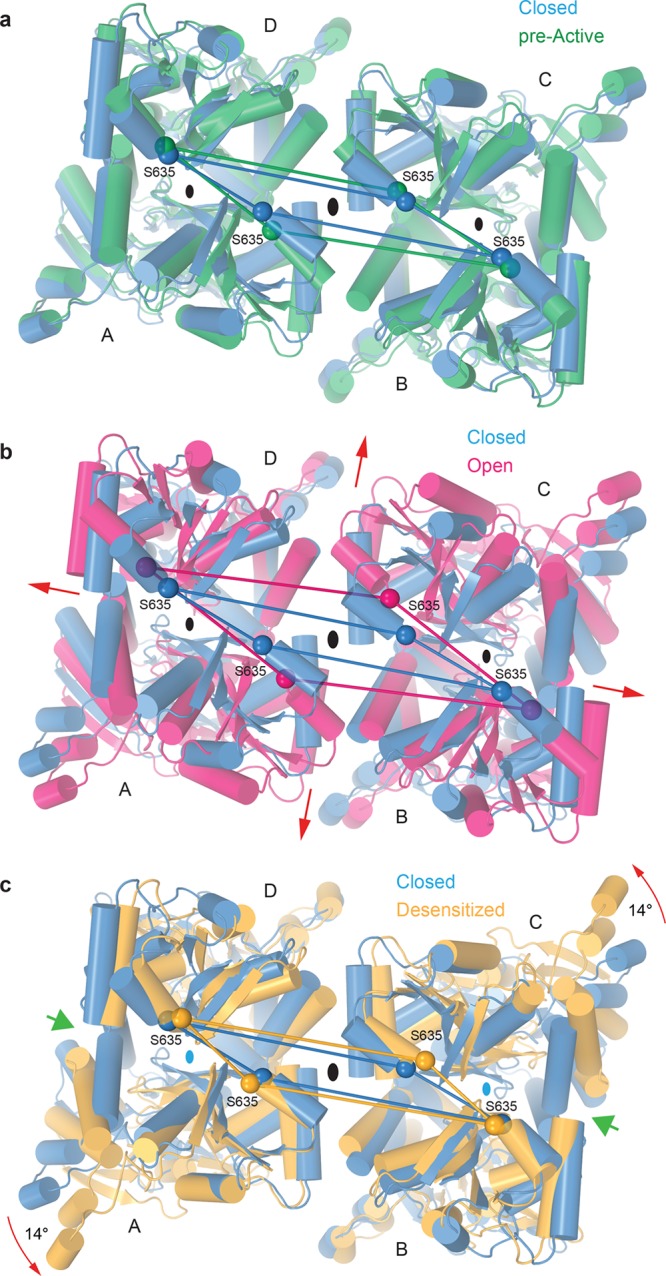
Structural rearrangements in the LBD tetramer during gating. Superposition of LBD tetramers from GluA2–GSG1LZK-1 (blue, closed state) and (a) GluA2NOW (green, pre-active state), (b) GluA2–STZGlu+CTZ (magenta, open state), or (c) GluA2–GSG1LQuis (orange, desensitized state) viewed from the TMD along the axis of the overall 2-fold receptor symmetry (large black ovals in the middle). Cα atoms of S635 are shown as spheres of the corresponding color, connected by straight lines. Broadening of the LBD layer in the open state and rotation of the A and C monomers in the desensitized state are indicated by red arrows. Green arrows in panel c point to the cleft between the desensitized state LBD protomers, signifying the loss of local 2-fold symmetry in LBD dimers (small ovals) and 3-fold smaller intradimer interfaces.
