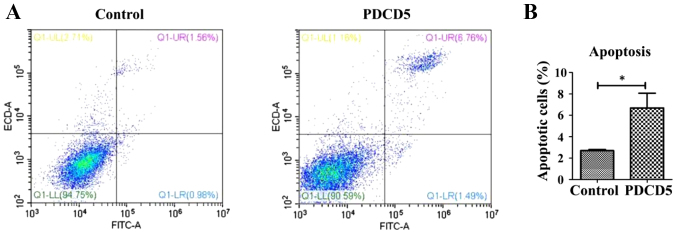
Figure 4.
Overexpression of PDCD5 induces apoptosis in A431 cells. Flow cytometry was performed to determine apoptosis in the PDCD5 overexpressing cells and its control cells. (A) More apoptotic cells were clearly observed in the PDCD5 overexpressing cells compared with the control cells. Representative images from three independent experiments are shown. (B) Quantification for the percentage of apoptosis in the PDCD5 overexpressing A431 cells and its control cells from three independent experiments. *P<0.05. Control, empty vector-transfected A431 cells; PDCD5, PDCD5 overexpressing A431 cells.

