Figure 4.
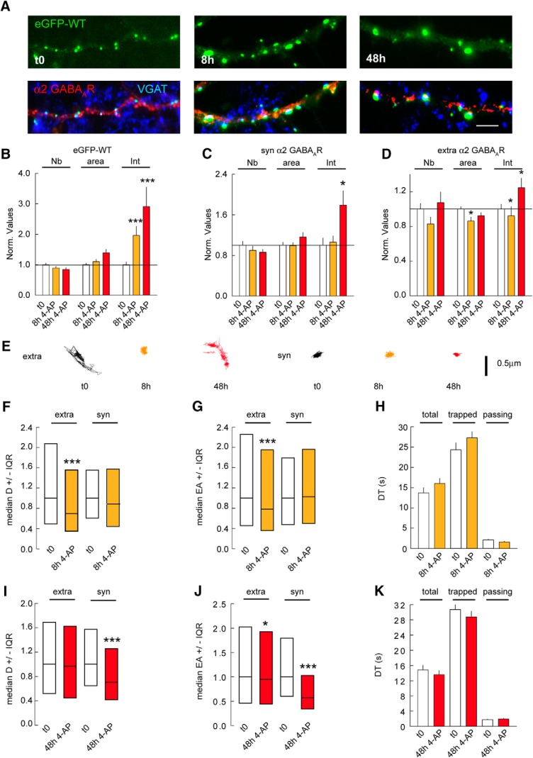
Gephyrin clustering influences GABAAR lateral diffusion. A, Morphology of eGFP-WT (green) after 8 and 48 h of 4-AP application; VGAT (blue), GABAAR α2 (red) at 21 DIV. Scale bar, 10 µm. B, Quantification of eGFP-WT clusters after 8 and 48 h of 4-AP application. t0 n = 55 cells, 8 h n = 46 cells, 48 h n = 55 cells, 3 cultures. Cluster Nb: 0–8 h: p = 0.13, 0–48 h: p = 0.002; Area: 0–8 h: p = 0.5, 0–48 h: p = 0.001; Intensity: 0–8 h: p < 0.001, 0–48 h: p < 0.001. C, Quantification of synaptic α2 GABAAR clusters after 8 and 48 h of 4-AP compared with mock treated control. t0 n = 52 cells, 8 h n = 43 cells, 48 h n = 53 cells, 3 cultures. Cluster Nb: 0–8 h: p = 0.4, 0–48 h: p = 0.3; Area: 0–8 h: p = 0.8, 0–48 h: p = 0.8; Intensity: 0–8 h: p = 0.5, 0–48 h: p = 0.03. D, Quantification of extrasynaptic α2 GABAAR clusters after 8 and 48 h of 4-AP compared with mock treated control. t0 n = 52 cells, 8 h n = 43 cells, 48 h n = 53 cells, 3 cultures. Cluster Nb: 0–8 h: p = 0.2, 0–48 h: p = 0.9; Area: 0–8 h: p = 0.02, 0–48 h: p = 0.3; Intensity: 0–8 h: p = 0.05, 0–48 h: p = 0.022. E, Example trace of α2 GABAAR trajectories showing surface exploration of extrasynaptic and synaptic receptors after 8 and 48 h of 4-AP exposure. Scale bar, 0.5 µm. F, Quantification of diffusion coefficients of α2 GABAAR after 8 h of 4-AP exposure. Extra; t0 n = 450 QDs, WT 4AP 8 h n = 961 QDs, p = 1.96 10−7. Syn; t0 n = 103 QDs, 8 h n = 138 QDs, p = 0.22; 2 cultures. G, Quantification of explored area EA of α2 GABAAR after 8 h of 4-AP application. Extra; t0 n = 1347 QDs, 8 h n = 5265 QDs, p = 6.4 × 10⁻⁹. Syn; t0 n = 308 QDs, 8 h n = 708 QDs, p = 0.63. H, Quantification of synaptic dwell time DT of α2 GABAAR showing no impact after 8 h of 4-AP for total, trapped, or passing receptor population. Total: t0 n = 151 QDs, 8 h n = 206 QDs, p = 0.073; Trapped: t0 n = 80 QDs, 8 h n = 116 QDs, p = 0.36; Passing: t0 n = 78 QDs, 8 h n = 90 QDs, p = 0.02. I, Quantification of diffusion coefficients of α2 GABAAR after 48 h of 4-AP application. Extra: t0 n = 777 QDs, 48 h n = 174 QDs, p = 0.69. Syn: t0 n = 126 QDs, 48 h n = 213 QDs, p = 1.4 × 10⁻⁴. J, Quantification of explored area EA of α2 GABAAR after 48 h of 4-AP application. Extra: t0 n = 2331 QDs, 48 h n = 5508 QDs, p = 0.045. Syn: t0 n = 378 QDs, 48 h n = 717 QDs, p = 2.2 × 10⁻²⁰. K, Quantification of α2 GABAAR dwell time after 48 h of 4-AP application. Total: t0 n = 201 QDs, 48 h n = 254 QDs, p = 0.74. Trapped: t0 n = 91 QDs, 48 h n = 110 QDs, p = 0.99. Passing: t0 n = 110 QDs, 48 h n = 144 QDs, p = 0.81. In B–D, H, and K, data are presented as mean ± SEM, *, p ≤ 0.05; ***, p ≤ 0.001 (Mann–Whitney rank sum test). In F, G, I, and J, data are presented as median values ± 25%–75% IQR, *, p ≤ 0.05; ***, p ≤ 0.001 (Kolmogorov–Smirnov test). In B–G, I, and J, values were normalized to the corresponding control values. In H and K, DT in s.

