Figure 1.
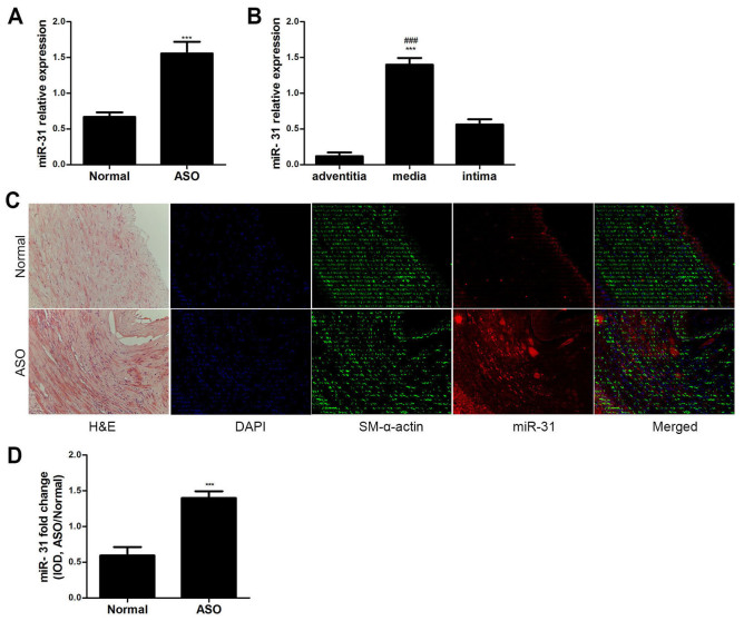
Characteristics of miR-31 expression. (A) miR-31 expression in both ASO and normal arteries was determined by RT-qPCR (n=6). ***P<0.001 vs. normal group. (B) miR-31 expression in the three layers of the ASO artery wall was determined by RT-qPCR (n=6). ***P<0.001 vs. adventitia and ###P<0.001 vs. intima. (C) H&E staining revealed artery structures. Co-staining of miR-31 (red) and SM-α-actin (green) in artery sections. The ISH and immunofluorescence results showed that miR-31 was primarily localized in the arterial media and neointima. (D) IOD value of miR-31 staining in artery sections showed that miR-31 staining was significantly increased in ASO sections (n=6) compared with that in normal sections (n=6). Magnification, ×200. ***P<0.001 vs. normal group. miR, microRNA; ASO, arteriosclerosis obliterans; RT-qPCR; H&E, hematoxylin and eosin; SM, smooth muscle; IOD, integrated optical density; DAPI, 4′,6-diamidino-2-phenylindole; ISH, in situ hybridization.

