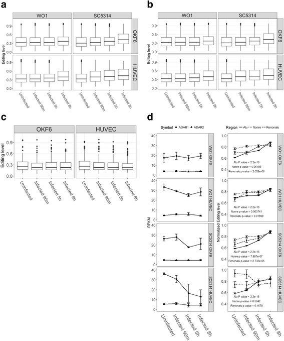
Fig. 2.

Pattern of RNA editing during the course of infection in epithelial and endothelial cells. a Distribution of editing level in different infection conditions. b Distribution of editing level in stable expression genes. c Distribution of editing level in significant differential expression genes. d Pattern of ADAR genes expression and the normalized editing level of common editing site
