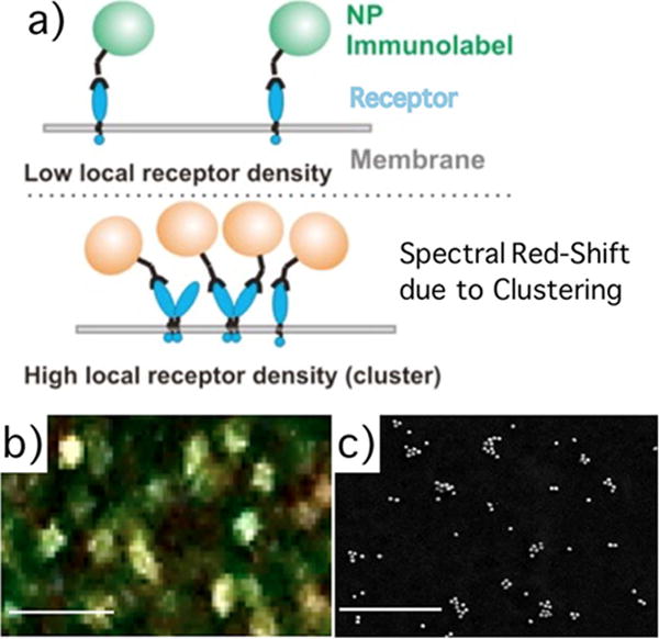
Figure 15.

(a) Concept of plasmon coupling microscopy. Clustering of NPs bound to a target receptor results in spectral shifts. (b) Color image of 40 nm diameter gold NP targeted to CD44 surface proteins on a MCF7 cell. (c) SEM image of NP immunolabels targeted to CD24. Adapted with permission from [96]. Copyright (2013) American Chemical Society.
