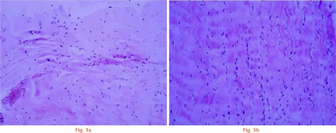
Pathological changes in the tissues in group A and B rabbits were compared after haematoxylin and eosin staining (x100). Compared with group A, the collagen fibre in the ligament tissue of group B rabbits formed a wave-like structure with no broken collagen fibres. Fibroblasts increased in number, but there were no visible changes in the nucleus. Fibroblasts exhibited a regular oval shape and were evenly distributed, which represents a recovered ligament tissue.

An official website of the United States government
Here's how you know
Official websites use .gov
A
.gov website belongs to an official
government organization in the United States.
Secure .gov websites use HTTPS
A lock (
) or https:// means you've safely
connected to the .gov website. Share sensitive
information only on official, secure websites.
