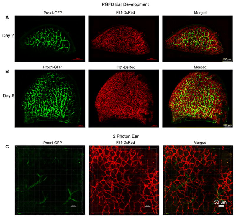
Fig. 1. Confocal microscopy and two-photon images of blood and lymphatic vessels in the PGFD mouse ears.

Ears harvested on postnatal days 2 and 6 and from adult mice were imaged using confocal microscopy. Scale bars: (A) 500 μm, (B) 500 μm, and (C) 50 μm.
