


Fig. 13. VEGFA–induced corneal HA and LA in tamoxifen-treated CDh5CreERT2/VEGRF2lox/PGFD mice.
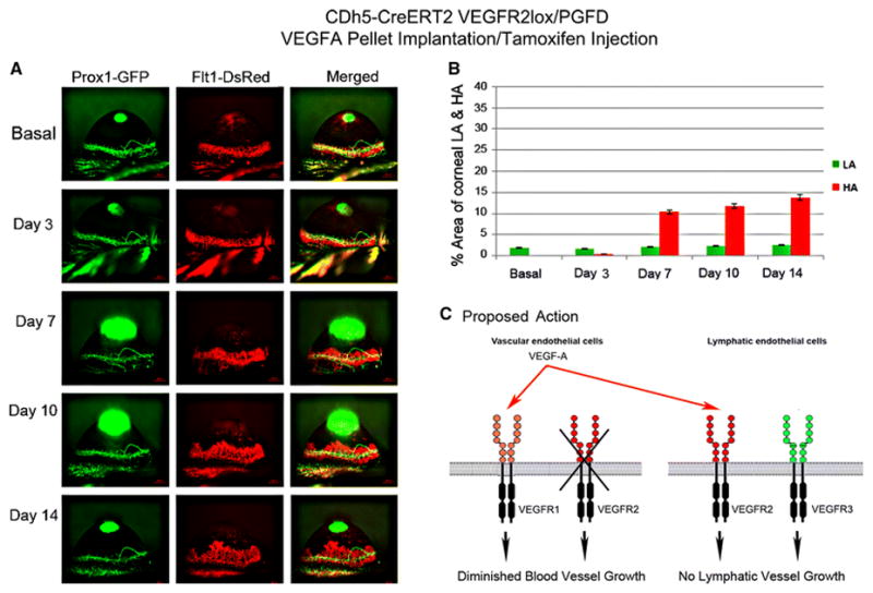
Axiozoom stereo microscopy was used to image the vascular (red) and lymphatic (green) vessels within the same cornea at each time point. (1A) Eliminated corneal LA and significantly increased HA in VEGFR2 knockout in vascular endothelial cells were observed on days 7, 10, and 14 after 150 ng VEGFA corneal implantation when compared to that observed in the tamoxifen-treated CDh5-CreERT2VEGRF2lox/PGFD mice. (1B) The percent area occupied by VEGFA–induced corneal HA and LA was calculated over the experimental time course.