Figure.

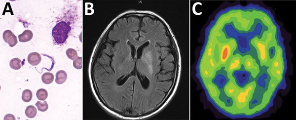
Bone marrow test results and brain imaging of a 60-year-old man who returned to China from Gabon with suspected human African trypanosomiasis. A) Trypanosoma spp. (later determined to be T. brucei gambiense) in a Giemsa-stained thin bone marrow film. Original magnification ×1,000. B) A T2-weighted fluid-attenuated inversion recovery image with hyperintense signal changes in the left basal ganglia. C) Brain positron emission tomography–computed tomography suggested reduced glucose metabolism in the left basal ganglia.
