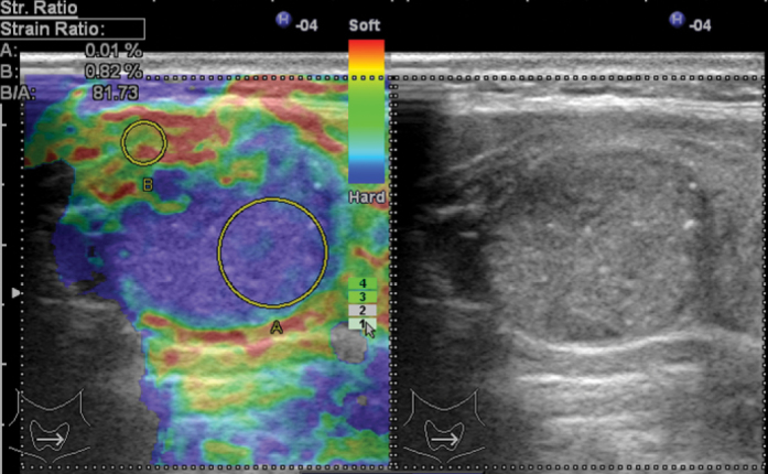
Figure 1.

Malignant nodule sample; Gray scale: iso-hypoechoic, including microcalcifications, Transverse/Longitudinal Axis ratio >0.5.
Elastography: Especially posterior region of nodule is observed as dark blue. Score 4. (Image Archive of Yıldırım Beyazıt University Faculty of Medicine, Department of Endocrinology and Metabolic Diseases).
