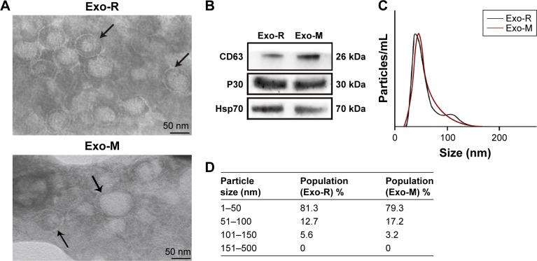
Figure 1.
Characterization of Toxoplasma gondii-derived exosomes.
Notes: (A) Representative TEM images of vesicles derived from T. gondii RH strain or ME49 strain showing a range of exosomal morphologies (original magnification, × 100,000; scale bar =50 nm). Bilayer membranes are arrowed. TEM was performed at least three times. (B) Western blotting analysis of T. gondii-derived vesicles with anti-CD63, anti-P30 (anti-SAG1) and Hsp70. (C) Size distribution of purified vesicles derived from T. gondii using nanoparticle tracking analysis and showing a mean diameter of 50 nm. (D) Table from NanoSight NS300 analysis of percentage of purified vesicles derived from T. gondii in various size ranges.
Abbreviations: Exo-R, exosomes from RH strain; Exo-M, exosomes from ME 49 strain; TEM, transmission electron microscopy.

