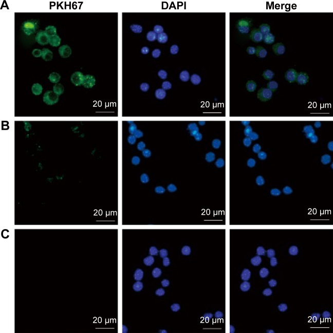
Figure 2.
Uptake of Toxoplasma gondii exosomes by macrophages.
Notes: Ten micrograms of the (A) PKH67-labelled T. gondii exosomes, or (B) PKH67-PBS control, or (C) PBS control alone were added to the macrophages and incubated at 37°C for 2 h. In the fluorescence microscopy pictures, PKH67 was used to label the exosomes (green) and DAPI was used to detect the nucleus of the macrophages (blue). Results are representative of three independent experiments.
Abbreviations: PBS, phosphate buffered saline; DAPI, 4′,6-diamidino-2-phenylindole.

