Figure 1.
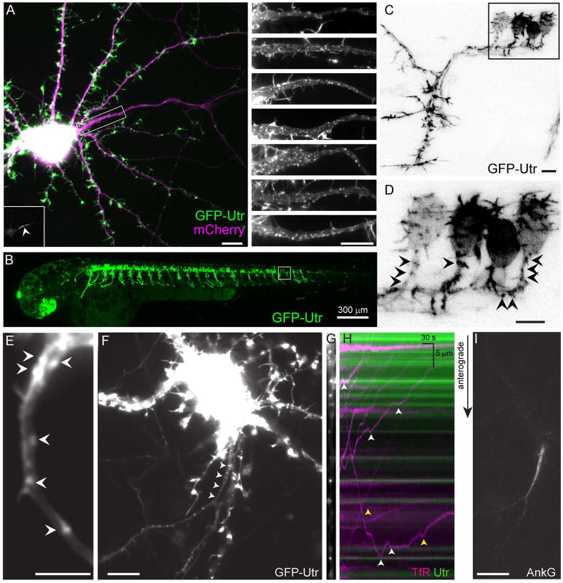
Actin patches are found in the proximal axons of live neurons and mark positions where vesicles carrying dendritic proteins halt and reverse. (A) 15 DIV cortical neuron in dissociated culture expressing the actin label GFP-Utr (green) and mCherry (purple). The proximal axon is contained within the boxed area. (Inset, lower left) Ankyrin G labeling of the proximal axon (arrowhead). (Right) Upper panel shows magnified image of boxed area. Lower panels show similar areas from other cortical neurons in culture expressing GFP-Utr under similar conditions. N = 20 neurons, 3 cultures. (B) Live 2 dpf zebrafish expressing GFP-Utr in motoneurons. N = 295 cells, 4 fish. (C, D) Higher magnification of area shown in boxed region of (B, C). (D) Arrowheads point to patches of actin in the proximal axon. (E) Proximal axon of 3 DIV cortical neuron expressing GFP-Utr showing actin patches (arrowheads). N = 15 neurons, 4 cultures. (F) 14 DIV cortical neuron in culture expressing TfR-mCherry (arrowheads point to the proximal axon). (G) Straightened image of proximal axon showing GFP-Utr labeling of actin patches. (H) Kymograph of vesicles carrying TfR-mCherry (purple) and actin patches (green). White arrowheads indicate places where TfR containing vesicles halted near actin patches, yellow arrowheads indicate places where halting took place away from patches. (I) Ankyrin G labeling of the axon initial segment of neuron in (F–H). Scale bar 10 µm unless otherwise indicated. See also Figures S1, S2, Movies S1, S2.

