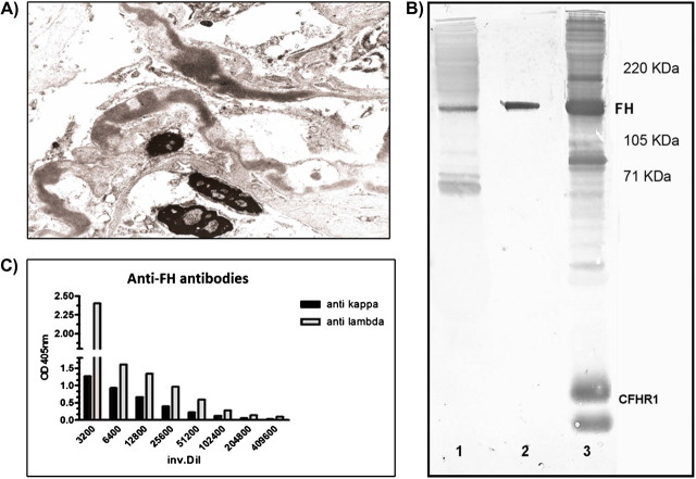
Fig. 1.
(A) High-power microscopic view shows the segmental irregular distribution of the ribbon-like thickenings of the glomerular basement membranes by intramembranous highly electron-dense deposits. (B) Western blot. SDS–PAGE carried out with IgG depleted normal human serum (NHS) in well 1 and 3 (corresponding to lane 1 and 3 in the figure) and with purified FH in well 2. After transfer, lanes 1 and 2 were revealed with IgG purified from the patient and lane 3 with a polyclonal anti-FH antibody that identifies FH and some Complement Factor H-related proteins (CFHR) in NHS. (C) Anti-FH antibody light chain isotype determination by ELISA assay. Anti kappa, anti-human κ light chain antibody; anti lambda, anti-human λ light chain antibody; inv. Dil, serum inversal dilution; CFHR1, complement Factor H-related protein 1.

