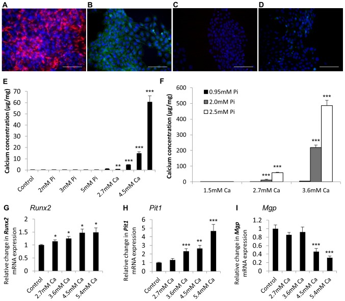
Figure 5.
Characterisation of RAVICs confirms the use of immortalised VICs as an in vitro CAVD model. Positive staining for (A) vimentin and (B) α-SMA in the rat aortic valve interstitial cell line (RAVIC). Representative images of (C) mouse and (D) rabbit IgG negative control. DAPI was used as a counterstain. Scale bar=100 µm. Calcium deposition determined by HCl leaching (µg/mg protein) in VICs treated with (E) calcium (Ca) alone (1.5–5.4 mM) and phosphate (Pi) alone (1.5–5.0 mM) and (F) calcium and phosphate in combination (1.5 to 3.6 mM Ca/0.95 to 2.5 mM Pi). Fold change in the mRNA expression of (G) Runx2 (H) PiT1 and (I) Mgp in VICs treated with Ca (2.7–5.4 mM) for 48 h. Results are presented as mean ± SEM. *P<0.05, **P<0.01; ***P<0.001 compared to control; n=6.

