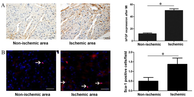
Figure 1.
bFGF protein expression and Sca-1+ CSC recruitment following myocardial infarction. (A) Immunohistochemistry analysis of bFGF protein expression in different areas of the heart one week following myocardial infarction. bFGF was stained brown and nuclei were stained blue. (B) Immunofluorescent staining of Sca-1+ CSC in different areas of the heart one week after myocardial infarction. Consecutive 10 sections and 10 random ×200 fields per section were counted. *P<0.05, with comparisons indicated by lines. bFGF, basic fibroblast growth factor; CSC, cardiac stem cells. Bar=100 µm.

