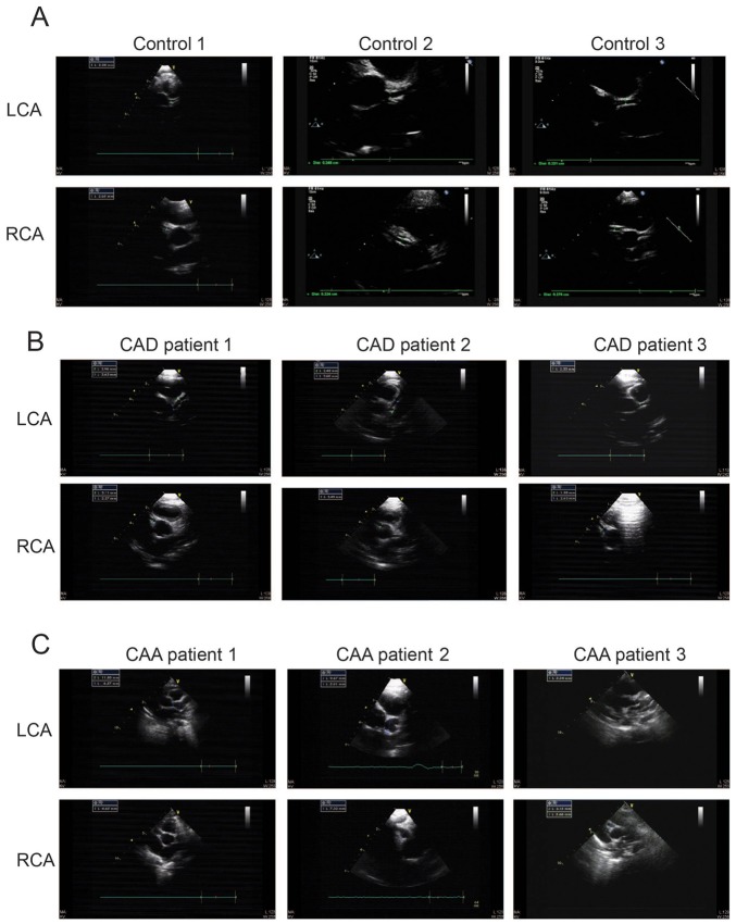
Figure 1.
ECG images of coronary arteries from healthy individuals and patients with Kawasaki disease. Representative ECG images from (A) three children with normal left and right coronary arteries (controls); (B) three patients with CAD; and (C) three patients with CAA. ECG, echocardiography; CAA, coronary artery aneurism; CAD, coronary artery dilation; LCA, left coronary artery; RCA, right coronary artery.

