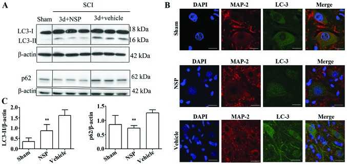
Figure 4.
NSP can restore disruption of autophagic flux after SCI. (A) Western blot analyses of LC3 and p62 protein in the spinal cord at 72 h after SCI. (B) Quantitative analysis of western blot analyses indicated that NSP reduces LC3-II and p62 protein levels at 72 h after SCI. Mean ± SD (n=6/group). *P<0.05, **P<0.01 compared with the vehicle-group. (C) Representative confocal images of LC3 and MAP-2 immunofluorescence double staining in the sham group, NSP-treated and vehicle-treated animals. Vehicle-treated rats exhibited increased dots of LC3 compared with the sham control at 72 h after SCI. NSP decreased the dots of LC3 vs. vehicle control (n=3/group; scale bars, 50 µm).

