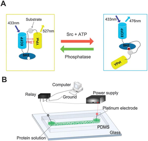
Fig. 1.

Schematic of the ECFP/YPet Src biosensor and its delivery by electroporation. (A) The mechanism of the Src FRET biosensor. The FRET signal varies with the Src activity and phosphatase treatment. (B) The setup for electroporation-based biosensor delivery in a microfluidic channel. A microfluidic channel facilitates applications of electric pulses of milliseconds and the observation of cellular dynamics. The dimensions of the channel were 150 μm (W) × 40 μm (D) × 3.8 mm (L).
