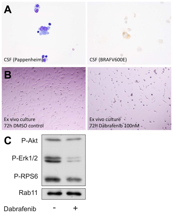
Figure 5.
Analyses of cerebrospinal fluid (CSF) and ex vivo tumor cell culture. Pappenheim staining of CSF cells (A, left) and immunohistochemistry for BRAF V600E (A, right) is shown. The lowest panel shows light microscopic images of the ex vivo tumor cell culture after 72 h of treatment with DMSO control and the BRAF V600E inhibitor dabrafenib. (B) Microscopic photographs of the cells 72 h after treatment with DMSO control (left) and after treatment with dabrafenib at a concentration of 100 nM (right). Western blot analysis is shown in (C). Phosphorylation of ERK is inhibited by dabrafenib.

