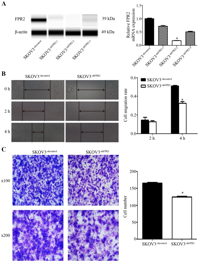
Figure 4.
(A) Validation of FPR2 expression in FPR2-knockdown SKOV3 cells using RT-qPCR and Simple Western assays. The mRNA and protein levels of FPR2 were significantly decreased in the SKOV3−shFPR2 cells compared with the control cells, with the SKOV3−shFPR2-2 cells exhibiting the greatest inhibition. (B) The wound healing assay showed that FPR2 knockdown significantly decreased the cell migration rate at 4 h after scraping. Significantly different from the control (SKOV3−shcontrol; *P<0.05). (C) The Transwell assay showed that FPR2 knockdown decreased the number of invasive SKOV3 cells (*P<0.05).

