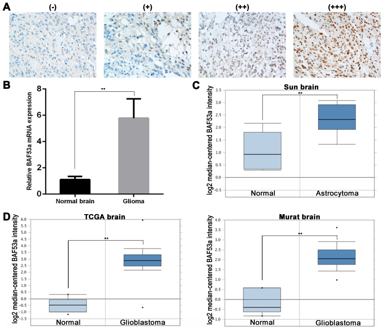
Figure 1.
BAF53a expression is upregulated in glioma. (A) The representative images of BAF53a expression is detected by IHC in glioma tissues (scored as - to +++). Magnification, ×400. (B) The real-time PCR results show that the expression level of BAF53a mRNA in glioma tissues was higher than that in normal brain tissues. The expression profile of BAF53a in Oncomine database: BAF53a gene copy number in astrocytoma is higher than normal brain tissues in Sun brain data (C), and BAF53a gene copy number in glioblastomas is significantly higher than normal brain tissues in TCGA brain data and Murat brain data (D). *P<0.05; **P<0.01.

