Figure 5.
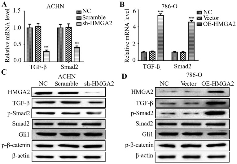
Silencing of HMGA2 decreased TGF-β and Smad2 expression in renal cell carcinoma cells. qPCR was used to detect the expression of TGF-β, Smad2 and β-actin in HMGA2-depleted ACHN (A) and HMGA2-overexpressing 786-O cells (B). Quantification from three independent experiments is shown as mean ± standard deviation (SD). ***P<0.001 and ****P<0.0001. Western blotting was used to detect the protein levels of HMGA2, TGF-β, phosphorylated-Smad2 (p-Smad2), Smad2, Gli1, phosphorylated-β-catenin (p-β-catenin) and β-actin in HMGA2-depleted ACHN cells (C) and HMGA2-overexpressing 786-O cells (D). Representative protein bands from three experiments are shown.

