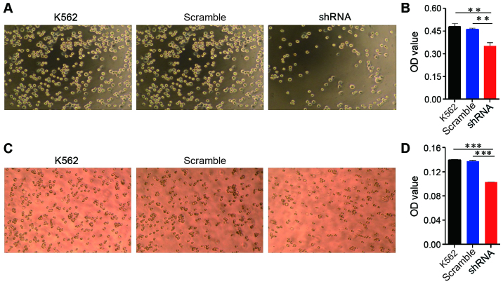
Figure 3.
EPS8 increases the adhesion and migration of leukemia cells. (A and B) K562 cells and their derivatives (1×10⁵ cells/well) were suspended in RPMI-1640 with 10% FBS and then added to a fibronectin-coated 96-well plate, incubated for 1.5 h and washed gently. The cells that adhered to the bottom of the plate were observed under a microscope. (A) The representative plots of the indicated strains under the microscope. (B) The number of adhered cells was assessed using the CCK-8 assay. (C and D) Transwell chambers were used to assess migration. Cells ~1×10⁵ were placed on the upper layer of a cell permeable membrane in serum-free RPMI-1640 containing 0.1% bovine serum albumin and allowed to migrate to the lower chamber, which contained RPMI-1640 and 10% FBS, for 6–8 h at 37°C. The cells that had migrated to the lower chamber were observed under a microscope. (C) The representative plots of the indicated strains under the microscope. (D) The number of migrating cells was calculated using the CCK-8 assay. Plots are representative of 3 independent experiments. **P<0.01 vs. the control; ***P<0.001 vs. the control.

