Figure 3.
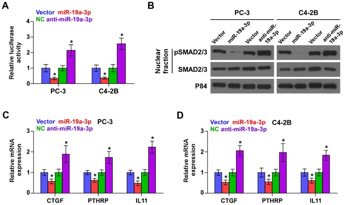
Downregulation of miR-19a-3p activates TGF-β signaling in PCa cells. (A) Transcriptional activity based on a TGF-β/Smad-responsive luciferase reporter as assessed in the indicated cells. Error bars represent the mean ± SD of three independent experiments. *P<0.05. (B) Western blot analysis showing that upregulation of miR-19a-3p decreased, while downregulation of miR-19a-3p increased nuclear translocation of pSMAD2/3 in PCa cells. The nuclear protein p84 was used as a nuclear protein marker. (C and D) Real-time PCR analysis of downstream bone metastasis-related genes of the TGF-β pathway, including CTGF, PTHRP and IL-11 in the indicated cells. Transcript levels were normalized to GAPDH expression. Error bars represent the mean ± SD of three independent experiments. *P<0.05.

