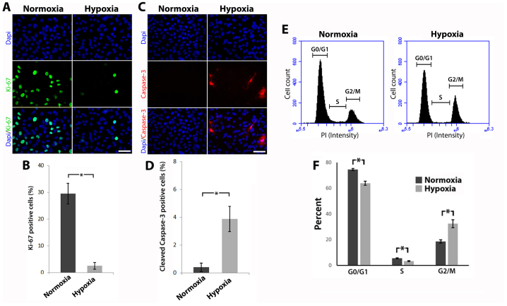
Figure 2.
Hypoxia reduces proliferation, perturbs cell cycle progression and induces apoptosis. (A) Cells were labeled for Ki-67 using fluorescent immunohistochemistry (scale bar, 25 µm). (B) Percentage of Ki-67 positive cells (error bars ± standard deviation, *P≤0.05, Mann-Whitney U test, n≥3). (C) Cells were labeled for cleaved caspase-3 using fluorescent immunohistochemistry (scale bar, 25 µm). (D) Percentage of cleaved caspase-3 positive cells (error bars ± standard deviation, *P≤0.05, Mann-Whitney U test, n≥3). (E) Cell cycle distribution was evaluated using flow cytometry. (F) Graph displays the cell cycle phase expressed as a percentage of total cells (error bars ± standard deviation, *P≤0.05, Mann-Whitney U test, n≥3).

