Figure 4.
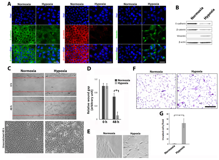
Hypoxia induces EMT, increased migration and invasion. (A) Cells were labeled for E-cadherin, β-catenin and vimentin using fluorescent immunohistochemistry (scale bar, 25 µm). (B) The levels of protein expression of E-cadherin, β-catenin and vimentin were detected by western blotting, β-actin served as a loading control. (C) Scratch wound migration assays were performed on confluent cells. Red dotted lines indicate the wound borders at the beginning of the assay. Lower panel displays comparative unscratched area. (D) Relative wound gap calculated as a ratio of the remaining wound gap at 48 h and the original wound gap at 0 h (error bars ± standard deviation, *P≤0.05; Mann-Whitney U test, n≥3). (E) Phase contrast images of migratory leading edge. (F) Photomicrographs of invaded cells in Matrigel Transwell assay. (G) Average number of invaded cells/field of view (error bars ± standard deviation, *P≤0.05; Mann-Whitney U test, n≥3).

