
Figure 7.
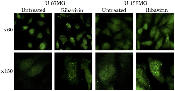
Observation of DSBs by γH2AX fluorescence microscopy. The accumulation of γH2AX in the cell nuclei was confirmed by fluorescence microscopy at 4 h following ribavirin treatment in the U-87MG and U-139MG cell lines.
Official websites use .gov
A
.gov website belongs to an official
government organization in the United States.
Secure .gov websites use HTTPS
A lock (
) or https:// means you've safely
connected to the .gov website. Share sensitive
information only on official, secure websites.
Observation of DSBs by γH2AX fluorescence microscopy. The accumulation of γH2AX in the cell nuclei was confirmed by fluorescence microscopy at 4 h following ribavirin treatment in the U-87MG and U-139MG cell lines.