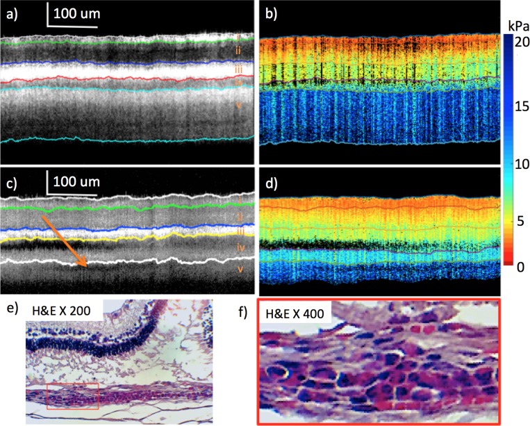
Figure 4.
Healthy versus abnormal rabbit from weeks 4 and 8 imaging after light treatment. (a) OCT of healthy retina at week 4. (b) Elastogram of healthy retina at week 4. (c) OCT of abnormal portion of retina at week 8. (d) Elastogram of abnormal portion of retina at week 8. (e) H&E staining after euthanization at week 8. (f) Higher magnification, H&E histology. Red box includes photoreceptor/RPE debris with round cell accumulation in underlying choroid and presumed lymphocyte filtration. Orange arrow (c) points to presumed lymphocytic infiltrate in choroid, resulting in low OCT signal.

