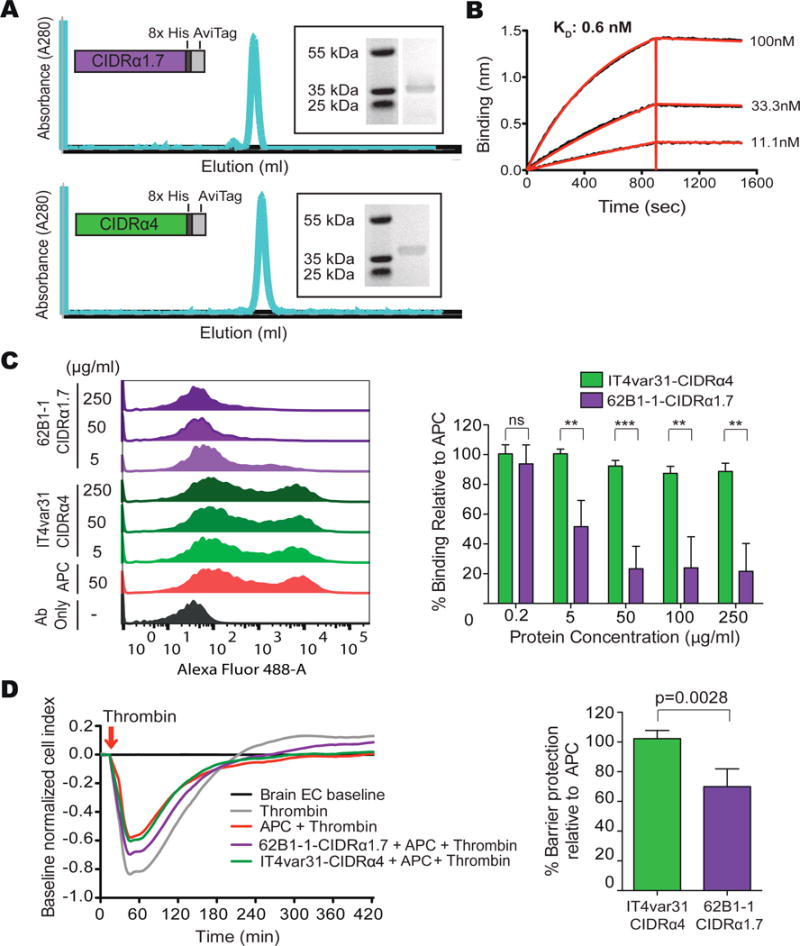
Figure 6. Inhibition of the APC-EPCR interaction by a common brain-specific 62B1-1-CIDRα1.7 domain isolated from a fatal pediatric CM case.

(A) Schematic of the expressed recombinant proteins, size exclusion chromatography trace and SDS-PAGE.
(B) 62B1-1-CIDRα1.7 binds to EPCR with picomolar affinity. A representative graph from n = 3 independent experiments.
(C) 62B1-1-CIDRα1.7 blocks binding of APC to EPCR. Left: representative flow cytometry histograms show binding of APC to CHO745-EPCR. Right: percentage of APC binding to CHO745-EPCR cells in the presence of varying concentrations of the CIDR domains, relative to APC binding alone. Mean ± SD of n = 3–5 independent experiments. Ab: Antibody. See Figure S5.
(D) 62B1-1-CIDRα1.7 partially blocks APC-mediated protection from thrombin-induced barrier disruption of primary human brain endothelial cells (HBMECs). Left: graph showing the cell index over time. Right: graph showing the level of APC protection from thrombin-induced barrier disruption in the presence of 62B1-1-CIDRα1.7 and IT4var31-CIDRα4, relative to APC alone. Mean ± SD of n = 4 independent experiments **p<0.01, ***p<0.001 by the Student’s unpaired, two-tailed t-test.
