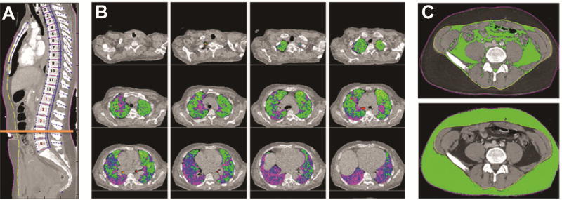
Figure 1.
Morphomic analysis using standard chest CTs showing (A) anatomic indexing of the vertebral levels to create an anatomical coordinate system, (B) lung density (LD) measurements where LD3 (green) to LD5 (purple) represent areas of increasing density, and (C) visceral and subcutaneous fat area after delineation of skin (purple) and fascia (yellow).

