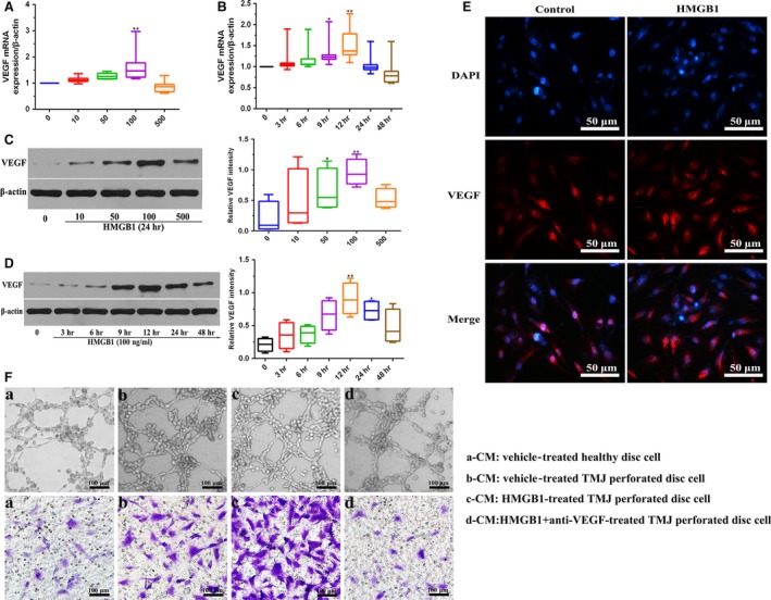
Figure 1.

Effects of HMGB1 on VEGF expression at the mRNA and protein levels in perforated disc cells of human TMJ. Perforated disc cells were stimulated with HMGB1 for various concentrations (0–500 ng/ml), mRNA and protein expression levels of VEGF in these cells were detected by qRT‐PCR (A) and Western blot (C). β‐actin served as an internal control. (B, D) qRT‐PCR and Western blot analysis of VEGF in these cells after treatment with HMGB1 (100 ng/ml). β‐actin served as an internal control. (E) Immunofluorescent staining of VEGF in these cells with or without HMGB1 (100 ng/ml) treatment for 24 hrs. Scale bar = 50 μm. (F) Different conditioned medium (CM) (a, b, c, d) was collected for tube formation assay and migration of HUVECs. Scale bar = 100 μm. Box plots, 25th and 75th percentiles; horizontal solid lines, medians; horizontal bars, minimum and maximum. *P < 0.05 and **P < 0.01 compared to the group without HMGB1 stimulation.
