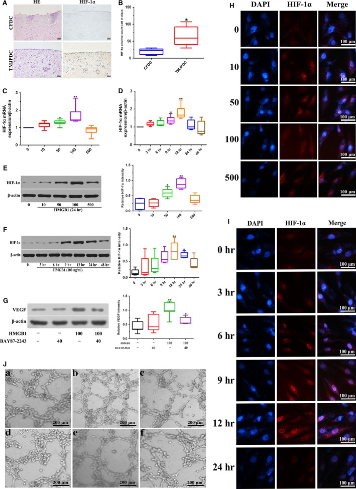
Figure 2.

Effects of HMGB1 on expression and activation of HIF‐1α in perforated disc cells of human TMJ. (A, B) Disc tissues from patients with disc perforation and from patients with condylar fracture were compared using HE, immunohistochemical staining against HIF‐1α, and the number of HIF‐1α‐positive staining cells was analysed. (C, E) Perforated disc cells were treated with different doses of HMGB1 (0–500 ng/ml) for HIF‐1α mRNA and protein analysis at 24 hrs. β‐actin was used as a loading control. (D, F) These cells were incubated for various times (0–48 hrs) with HMGB1 (100 ng/ml). HIF‐1α mRNA and protein levels were determined by RT‐PCR or Western blotting, respectively. β‐actin was used as a loading control. (G) Perforated disc cells were pre‐treated with BAY87‐2243 (40 μM) for 1 hr and then incubated HMGB1 (100 ng/ml) for 24 hrs. VEGF protein levels were determined by Western blotting. β‐actin was used as a loading control. (H, I) Effect of HMGB1 on activity of HIF‐1α in perforated disc cells using immunofluorescence. Scale bar = 50 μm. (J) Perforated disc cells were pre‐treated with BAY87‐2243 (40 μM), U0126 (60 μM), SB203580 (40 μM) or SP600125 (60 μM) for 30 min, and then incubated with HMGB1 (100 ng/ml) for 24 hrs. Culture medium was then collected for tube formation of HUVECs. Box plots, 25th and 75th percentiles; horizontal solid lines, medians; horizontal bars, minimum and maximum.*P < 0.05 and **P < 0.01 compared to the group without HMGB1 stimulation. # P < 0.05 compared to the group treated with HMGB1 alone. a. Conditioned medium (CM) from vehicle‐treated perforated disc cells; b. CM from HMGB1‐treated perforated disc cells; c. CM from HMGB1 + BAY87‐2243‐treated perforated disc cells; d. CM from HMGB1 + U0126‐treated perforated disc cells; e. CM from HMGB1 + SB203580‐treated perforated disc cells; f. CM from HMGB1 + SP600125‐treated perforated disc cells.
