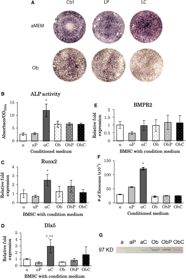
Figure 3.

Analysis of conditioned‐medium after LC treatment of BMSC culture. (A) ALP staining of BMSCs cultured in conditioned media for 1 week. Conditioned‐medium was made in α‐MEM or osteogenic medium (Ob) containing either LP or LC (0.1 mg/ml). The control did not contain liposomes. (B) ALP activity analysis in α‐MEM (α), LP‐conditioned‐medium (αP), LC‐conditioned medium (αC), osteogenic medium (Ob), LP‐conditioned osteogenic‐medium (ObP), and LC‐conditioned osteogenic‐medium (ObC). *: significantly different among all treatments (P < 0.05, n = 3). (C–E) Real time RT‐PCR of BMSCs cultured in conditioned‐media. mRNA expressions of Runx2 (C), Dlx5 (D) and BMPR2 (E) were examined (n = 3). (C) *: αC versus α, αP and ObC (P < 0.05); (D) *: αC versus α and ObC (P < 0.05); **: αC versus αP, Ob and ObP (P < 0.01). (F) The number of exosomes found in conditioned‐media. *: significantly different with all treatments (P < 0.05, n = 5). (G) Detection of RANK using western blot analysis in conditioned‐media. The size for RANK is 97 KDa. All statistical data are represented as mean ± SD by one‐way anova.
