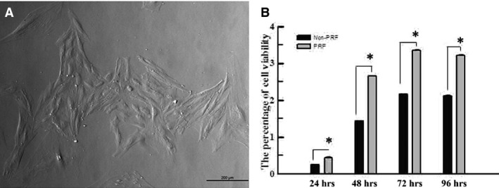
Figure 1.

(A) The colonies of rat PDLSCs could be observed microscopically, and cells in good condition exhibited a fusiform shape with an oval nucleus, lying in the middle of the cytoplasm (40×). (B) MTT assay indicated that the proliferation level of rat PDLSCs in PRF group was higher than that in normal group at 24, 48, 72 and 96 hrs in vitro. *Statistically significant difference compared to control and PRF group (P < 0.05).
